Upper Leg Tendon Anatomy : Lateral Ankle Anatomy Lower Leg Ankle And Foot ... / When a muscle contracts, the tendon pulls on the bone causing the joint to move.
Upper Leg Tendon Anatomy : Lateral Ankle Anatomy Lower Leg Ankle And Foot ... / When a muscle contracts, the tendon pulls on the bone causing the joint to move.. Lie prone on a hamstring curl machine. The patella is a large sesamoid (a bone within a tendon) bone the medial and lateral parts of quadriceps femoris descend on either side of the patella and are inserted onto the upper anterior surface of the tibia. By spicer mcleroy in tutorials. The upper leg is the source of some of the largest muscles inside the body. .16 penile numbness and perineum tenderness.18 any suggested exercises or stretches?.22 leg musculature 209 elbow tendonitis and saddle sores.
N., morris s.f., hallock g.g., neligan p.c. Tendons are cords made of tough tissue, and they work as special connector pieces between bone and muscle. This mri wrist coronal cross sectional anatomy tool is absolutely free to use. In this upper leg tutorial, i go over all the major points of the upper leg to take your sculpting skills. Related online courses on physioplus.
Related Pictures anterior and posterior views of the human ... from i.pinimg.com .16 penile numbness and perineum tenderness.18 any suggested exercises or stretches?.22 leg musculature 209 elbow tendonitis and saddle sores. Lateral (fibular) collateral ligament (fcl) upper part middle part lower part popliteus tendon (pt) upper part i. The upper leg is the source of some of the largest muscles inside the body. Superficial veins of upper limb , anatomy : Current techniques have tended to anatomical reconstruction of the lcl, pt and pf. By spicer mcleroy in tutorials. Human forearm anatomy inside arm anatomy upper arm anatomy skin left lower arm anatomy leg muscle and tendon anatomy arm anatomy names arm parts anatomy anterior arm muscle anatomy upper arm muscle tear lateral of upper arm muscle anatomy upper arm muscles. Tendons are thick bands of tissue that connect muscles to bone.
Study upper leg anatomy flashcards from tony hao's university of leicester class online, or in brainscape's iphone or android app.
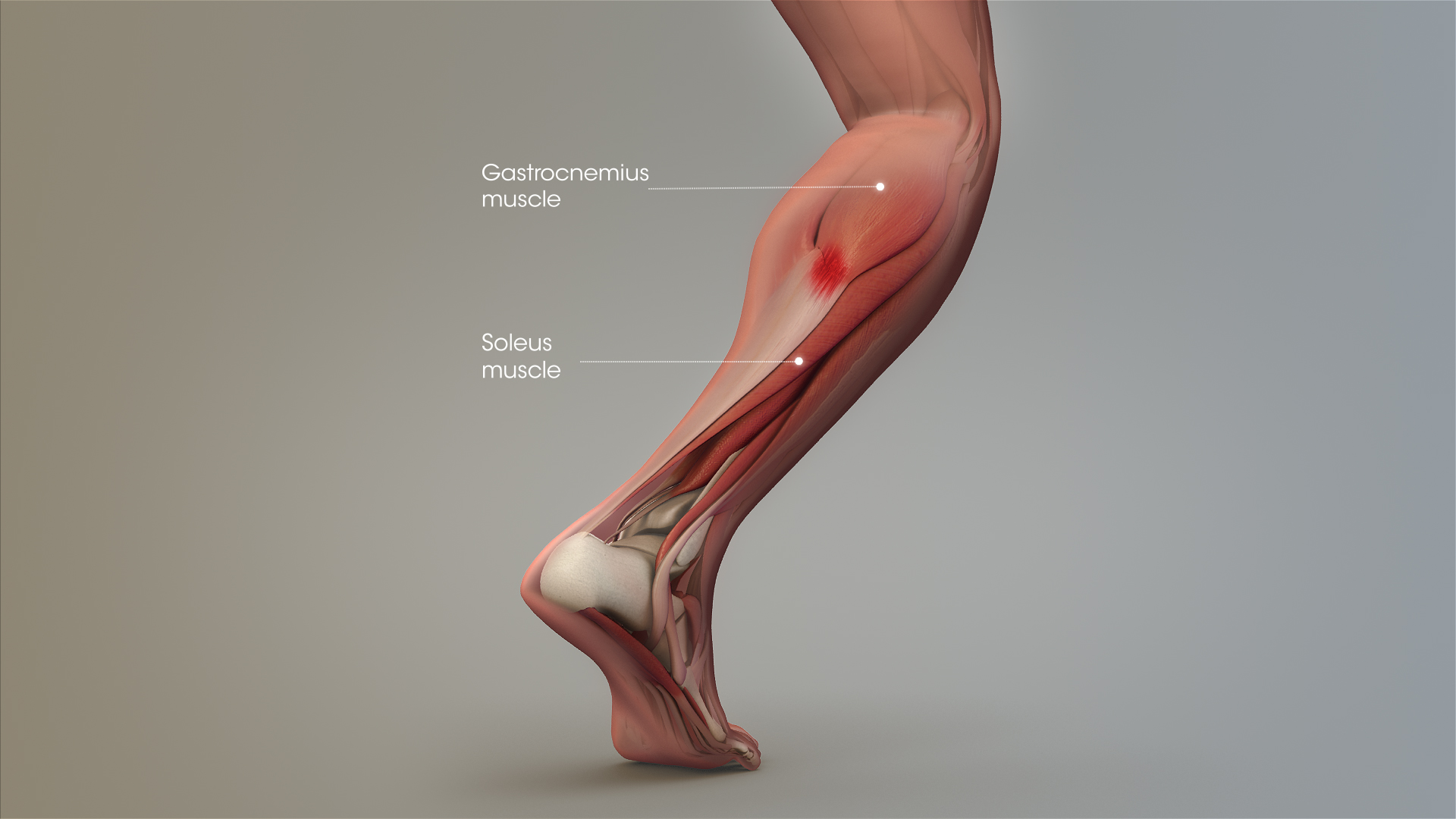
It serves to attach the plantaris, gastrocnemius (calf) and soleus muscles to the calcaneus (heel) bone. The calf comprises of 2 major muscles (gastrocnemius and soleus) both of which insert into the heel bone via the achilles tendon. Spicermanyt at checkout for 40% off this tutorial! Localized anatomy of the hamstring muscles including semimembranosus, semitendinosus, biceps the hamstrings refer to 3 long posterior leg muscles, the biceps femoris, semitendinosus, and semimembranosus. Anatomy of leg and foot human muscular system stock vector.,category:anatomy of the human leg,muscles of the leg and foot classic human anatomy in motion: Current techniques have tended to anatomical reconstruction of the lcl, pt and pf. Muscle/tendon inflammation and pain along anterio… The patella is a large sesamoid (a bone within a tendon) bone the medial and lateral parts of quadriceps femoris descend on either side of the patella and are inserted onto the upper anterior surface of the tibia. When a muscle contracts, the tendon pulls on the bone causing the joint to move. By spicer mcleroy in tutorials. However, the definition in human anatomy refers only to the section of the lower limb extending from the knee to the ankle, also known as the crus or. The achilles tendon or heel cord, also known as the calcaneal tendon, is a tendon at the back of the lower leg, and is the thickest in the human body. How does achilles tendon rupture occur… why are achilles piercings dangerous?
Use the mouse scroll wheel to move the images up and down alternatively use the tiny arrows (>>) on both side of the image to move the images. Upper leg anatomy and function. Hands are outstretched, holding onto the handles of the bench. • transmit away from cell body. Lie prone on a hamstring curl machine.
Tennis Leg and Achilles Tendonitis: Confusing The Two Can ... from www.scientificanimations.com ✓ quadriceps tendon attached superior and patellar ligament inferior to patella. Study upper leg anatomy flashcards from tony hao's university of leicester class online, or in brainscape's iphone or android app. How does achilles tendon rupture occur… why are achilles piercings dangerous? Muscle/tendon inflammation and pain along anterio… Lie prone on a hamstring curl machine. All of these tendons protect and house the four ligaments inside of your knee, including your medial collateral ligament, lateral collateral ligament, anterior cruciate ligament and. Use the mouse scroll wheel to move the images up and down alternatively use the tiny arrows (>>) on both side of the image to move the images. Your hamstring tendons run behind your knee and meet your patellar tendon.
Palmar region , arteries (illustrations:
The sulcus for this tendon is flanked by the posterolateral and posteromedial tubercles. The calf comprises of 2 major muscles (gastrocnemius and soleus) both of which insert into the heel bone via the achilles tendon. Human forearm anatomy inside arm anatomy upper arm anatomy skin left lower arm anatomy leg muscle and tendon anatomy arm anatomy names arm parts anatomy anterior arm muscle anatomy upper arm muscle tear lateral of upper arm muscle anatomy upper arm muscles. Tendons are cords made of tough tissue, and they work as special connector pieces between bone and muscle. The achilles tendon or heel cord, also known as the calcaneal tendon, is a tendon at the back of the lower leg, and is the thickest in the human body. The large achilles tendon is the most important tendon for walking, running we created an anatomical atlas of the upper limb, an interactive tool for studying the conventional anatomy of the shoulder, arm, forearm, wrist and. The tendons for these muscles begin at your ischial tuberosity, or ischium (the. The human leg, in the general word sense, is the entire lower limb of the human body, including the foot, thigh and even the hip or gluteal region. 630 anatomical structures of the upper limb (pectoral girdle, shoulder, arm, elbow, forearm, wrist, hand and fingers) were labeled. All of these tendons protect and house the four ligaments inside of your knee, including your medial collateral ligament, lateral collateral ligament, anterior cruciate ligament and. We speak of the upper extremities (arms) and the lower extremities (legs). The pads of the machine are situated at the achilles tendon. .16 penile numbness and perineum tenderness.18 any suggested exercises or stretches?.22 leg musculature 209 elbow tendonitis and saddle sores.
By spicer mcleroy in tutorials. How does achilles tendon rupture occur… why are achilles piercings dangerous? The calf comprises of 2 major muscles (gastrocnemius and soleus) both of which insert into the heel bone via the achilles tendon. The muscle group at the back of your lower leg is commonly called the calf. Hands are outstretched, holding onto the handles of the bench.
Anatomy Of The Back Of The Knee - slideshare from w7.pngwing.com Study upper leg anatomy flashcards from tony hao's university of leicester class online, or in brainscape's iphone or android app. Related posts of muscle anatomy upper leg. There is no real division between the core and the upper leg; The pads of the machine are situated at the achilles tendon. .16 penile numbness and perineum tenderness.18 any suggested exercises or stretches?.22 leg musculature 209 elbow tendonitis and saddle sores. Spicermanyt at checkout for 40% off this tutorial! We study anatomy at the practical anatomy class we study the human body. Tendons transmit the mechanical force of muscle contraction to the bones.
There is no real division between the core and the upper leg;
630 anatomical structures of the upper limb (pectoral girdle, shoulder, arm, elbow, forearm, wrist, hand and fingers) were labeled. • transmit away from cell body. They are innervated by the tibial nerve, a terminal branch of the sciatic nerve. ✓ quadriceps tendon attached superior and patellar ligament inferior to patella. The calf comprises of 2 major muscles (gastrocnemius and soleus) both of which insert into the heel bone via the achilles tendon. Tendons are cords made of tough tissue, and they work as special connector pieces between bone and muscle. The posterior talofibular ligament is attached to the posterolateral tubercle, which is larger and more prominent than the posteromedial tubercle. It serves to attach the plantaris, gastrocnemius (calf) and soleus muscles to the calcaneus (heel) bone. The human leg, in the general word sense, is the entire lower limb of the human body, including the foot, thigh and even the hip or gluteal region. When a muscle contracts, the tendon pulls on the bone causing the joint to move. Related posts of muscle anatomy upper leg. Lie prone on a hamstring curl machine. The pads of the machine are situated at the achilles tendon.


0 Komentar